Medical Imaging Projects for Research Scholars.
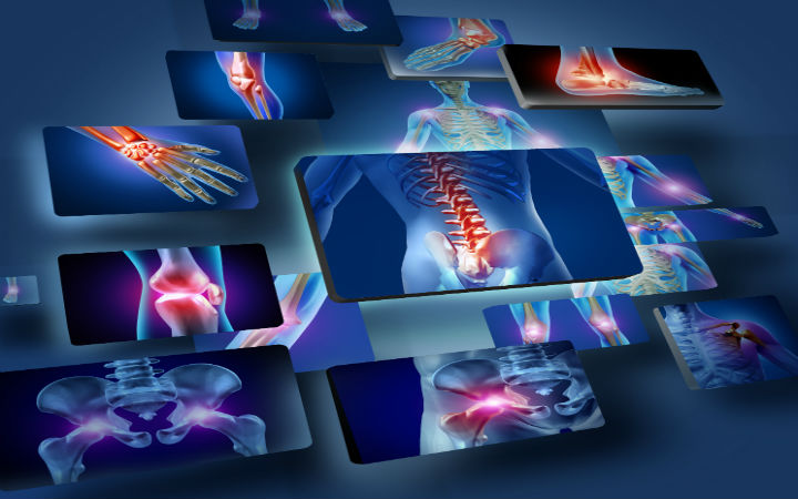
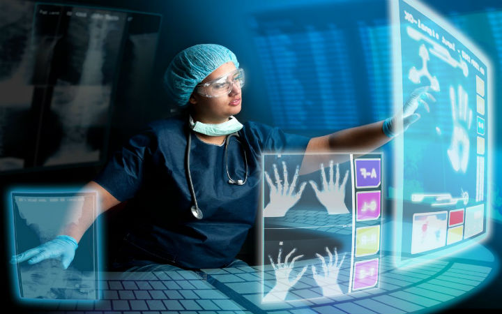
Medical imaging is otherwise known as diagnostic testing. Many medical images are taken in order to get a closer look at the disease. We offer Medical Imaging Projects for final year students and Research scholars. Medical images are taken in various diagnostic centers. Pre processing, segmentation, feature extraction, image enhancement, classification and feature reduction are the process that take place in medical imaging projects.Medical imaging is the technique and process of creating visual representations of the interior of a body for clinical analysis and medical intervention.
Types of Medical Imaging:
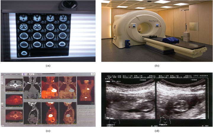
A biological action that takes place inside the body is understood by molecular imaging. It has the ability to detect the disease in its initial level. X- Ray beams are emitted inside the body. The beams spread at one part of the body and other part is taken up in x- Ray imaging. Ionizing radiation supports all medical imaging techniques. MRI and ultrasound imaging are the only medical imaging technique that does not require radiation.
Common advantages of different types of modalities:
X-Ray: Many injuries such as broken bones are detected using x-Ray.
PET: With little pain it can detect diseases in various body organs at the initial stage. It is expanded as positron emission tomography.
Ultrasound: Organs such as abdomen, kidneys, joints, muscles, bones, pelvis and breast are analyzed by ultrasound imaging to detect diseases. It is a painless and safe.
Computed Tomography: It is a fast and painless method. It has 3D volume facility and also spatial resolution recurrence of a disease or serious health issues can be detected using CT scan in Medical Imaging Projects.
MRI: It has the benefits of 3D acquisition, non-radiation and excellent contrast for all types of issues. MRI is used to get images of brain and knee.
Medical Imaging Projects.
Medical imaging is a process of making visual representations of the internal body parts and its functions. Some of the medical imaging projects are 3D reconstruction of blood vessels, semi-automated editing of medical images and spatial image fusion. Internal body behaviors can be gathered by the way of taking modalities of particular organs. Different types of modalities are presented in medical systems are CT, MRI and X-Ray. Human organ images are taken as an input and processed by different methods of feature extraction, segmentation and classification.
Knee segmentation has the processes of affine initialization, non-rigid registration (NRR), tissue classifier, bone segmentation and cartilage segmentation. Bone segmentation and cartilage segmentation methods are used to identify the healthy cartilage knee joints. Tongue analysis is also called as an important disease identification factor. To identify a Non proliferative Diabetic Retinopathy (NPDR) patients and healthy people we have to use feature based analysis process. Color, Texture and geometric features are extracted from tongue images and processed values can be used for identification process.
Medical Imaging Techniques:
Nuclear medical imaging shows how an organ to be functioned. It makes use of radioisotopes. It helps in the diagnosis of cancer and various organ disorders.
With certain properties of radiopharmaceuticals information regarding biochemistry and physiology can be obtained by nuclear medicine.
Enhance Imaging Projects:
It does the action of intensity transform which also includes processes like thresholding, logarithm transform, stretching and negative transform.
Medical image processing project was delivered to me on time .And the project was excellent. I am satisfied with the project
I would recommend academic project center to my friends as they are doing a great job.
Thanks for your guidance and support provided to me for completing my image processing project.
Very good faculty and experienced staff.They helped me to complete my project successfully.Thanks for your support.


