

Medical Image Processing Projects in Matlab lets gain closer details on human organs. Medical imaging is the emerging branch of medicine and technology. It has become an integral part of hospitals. Matlab simulation projects on medical image processing are very similar digital image processing. These projects can be done by students of information technology, electrical and instrumentation, computer science, biomedical engineering and electrical and instrumentation.
Advantages of Medical Image Processing Projects in Matlab:
- Image Guided Interventions and Minimally Invasive Surgery.
- Optimal Treatment Based On Personalized Medicine.
- Individual Risk Assessment.
- Screening the Specific Disease Entities.
- Early Detection of Subclinical Disease.
Overall Process of Medical Imaging: With the help of various modalities a human organ is scanned and taken for analysis. Output is obtained by processing many algorithms. This output is called as reconstructed cross sectional image.
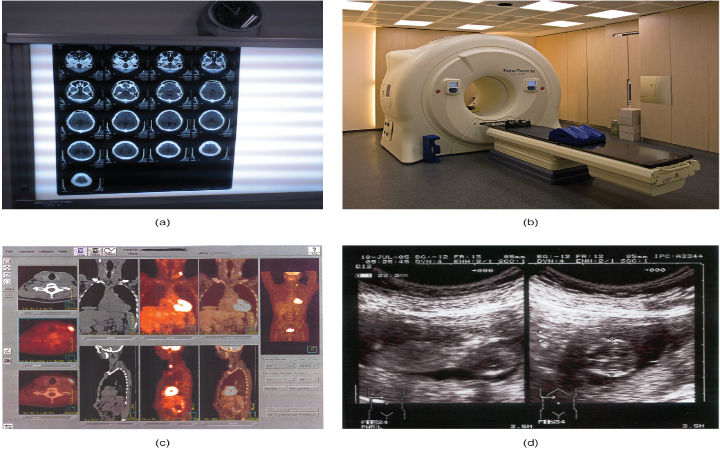
Image Modalities:
The modalities that are required to get the desired output are given below:
- PET/SPECT.
- MRI
- Computer Tomography (CT).
- X- Rays.
MRI: Magnetic field and radio frequency waves are used by MRI. It is otherwise known as effective imaging of protons. Hydrogen nucleus is the widely used image proton. Diseases such as brain position, tumor and disorder are detected using MRI.
Rays: Injuries on body parts, cells or bones can be detected using X- rays. Film of radiography used to take pictures. By x- rays electrons unit electromagnetic radiation.
Computer Tomography: It helps in detecting problems of head injury. It also helps in detecting lung cancer cells.Medical Image Processing Projects in matlab plays a vital role in computer tomography scans.
PET: Positron emission tomography aids doctors in identifying chemical or physiological changes that is related to metabolism. CT technique also helps in PET scans. Detection of sugar metabolism, blood flow, perfusion, oxygen utilization and receptor ligand binding rates.
Ultrasound: By using reflection of sound from the organ ultrasound pictures are taken. This sound information is then formed into an image.
Application and Types of Tomography: X-rays scans the whole body. Mammography scan is done by ultrasound technique. PET scans respiratory, brain and digestive systems. MRI can also perform whole body scan. Respiratory and digestive system is canned by radio isotopes.
Future Enhancement:
A wide range of topics focusing the field of disease identification is performed by researchers.
Medical Image Processing projects can be done by students of information technology, electrical and instrumentation, computer science, biomedical engineering and electrical and instrumentation. Projects on medical imaging lets get closer details on human organs.Medical imaging Processing Projects is the emerging branch of medicine and technology.
Medical Image Processing Projects
Digital image processing is a processing of scene data to be automatically processed by machine. Digital images are captured from digital cameras and scanners which have some pixel resolution, colors and image quality.Image analysis is a process of solving a problem of extracting information during manipulation of images in Image Processing Projects.
Digital Image Processing Projects
Image processing, computer vision and computer graphics are the categories of this field. Thesis should follow the sequence of problem formulation, creation of proposed algorithm, implementation and results. The manuscript of thesis should be published in a journal.Geoscience, remote sensing, medical imaging and signal processing are the promising areas of Image Processing thesis.
Image Processing Thesis


